Enhancing RIS-PACS solution with Artificial Intelligence

Enhancing RIS-PACS solution with Artificial Intelligence

It has become fairly clear of late that using technologies is aiding radiologists in not just enhancing patient care but also optimizing their time and effort. Several universities across the globe including the University of Virginia Health System are now looking for platforms that can be seamlessly integrated with Radiology Information System (RIS) and Picture Archival Communication System (PACS) to help not just streamline the workflow but also help detect findings that could be missed manually.

For instance, with the help of Artificial Intelligence (AI), loss in bone density can be found out at an early stage. There are probabilities of missing these detections while using the traditional interpretation method. Some AI Software uses colors to denote normal and abnormal findings. This speeds up the reading workflows in the PACS. Also, this is perfect for radiologists because then they know which results need his/her urgent attention. With AI software assisting in findings ranging from chest, pelvis CT scans and abdomen, coronary calcium, liver steatosis, pulmonary emphysema, spine compression fractures and bone mineral density, healthcare is all set to get transformed and how.

Preventive Care

An exciting step in preventive care, AI enhances RIS-PACS even as it leads to early detection of various conditions as well as ailments. It is but obvious that artificial intelligence is playing a key and an extremely vital role that could also go beyond regular readings and avert serious diseases from developing.

Sophisticated AI algorithms can now go hand-in-hand with RIS-PACS and deliver the added benefit of expediting the reading process while at the same time identifying findings that may go unobserved or are hard to visualize.

Enhanced Workflow

AI software can be running in the background and putting forth clinically significant and relevant findings that could have been missed.

Some software are developed with an access to in-house AI algorithms which integrates seamlessly with its RIS-PACS. This makes it convenient to easily integrate it with the workflow and give a unified AI experience to the user. These AI tools, when executed over the scanned images, empowers Radiologists to provide increased, consistently accurate and faster diagnosis.

Even in the sphere of Veterinary Sciences, AI helps with the workflow and enables integrated image management through central­ized scheduling to multiple connecting modalities, sites or centers. 

Automation Benefits

Automation is key for many AI tools and many radiologists prefer to call it the perfect assistant technology. Many AI software take on duties that make a radiologist’s job easier. When the radiologists are overburdened with studies, they tend to be in a rush and this in turn increases the error rates. AI puts together machine and humans and makes this combination much more powerful and error free.

Enhancing RIS-PACS, AI software works in tandem to lay emphasis and focus on high quality radiology reporting and accessibility. Deploying AI for radiology will only add immense value to patient diagnosis and care.

Eight point mantra for quality in teleradiology

Eight point mantra for quality in teleradiology

Addressing to what Dr Sona said in her article on Challenges in teleradiology in India,Dr Arjun Kalyanpur, MD, CEO and Chief Radiologist, Teleradiology Solutions, Bangalore shares his insights on quality in teleradiology reporting

Teleradiology has become firmly established as a powerful clinical paradigm within healthcare delivery that permits radiology reporting to be performed rapidly and efficiently as well as providing access to radiologic expertise where/when it might otherwise not have been available, in an era of crippling radiologist shortages. However, for teleradiology to provide continuing benefit, it must be supported by quality reporting, or else its value proposition ceases to exist. How can teleradiology providers ensure that they deliver consistent quality to their clientele, and through them to their patients? And how can a hospital or imaging center that is looking for a teleradiology provider decide which service really and truly represents a quality performer? Here are eight key processes that define quality in teleradiology, and can/will distinguish the quality teleradiology provider from the rest.

  1. Selection and training process

    An important part of the growth of a teleradiology practice as new radiologists are added to the team, is to adhere to stringent recruitment processes that include not just vetting the CV but also conducting a baseline reporting quality check. Given that teleradiology presents a wide variety of reporting challenges such a process ensures that the aspirations of the radiologist are matched with the quality standards and processes of the teleradiology organisation.

  2. Reporting standards and checklists

    It is necessary for a teleradiology provider to develop clear internal standards, checklists and reporting templates to ensure that quality is maintained in day-to-day reporting. These should be available for each modality, and ensure an internal standard of reporting that forms the bedrock of teleradiology reporting operations.

  3. Robust peer review

    At the heart of any successful teleradiology practice is a strong peer review process. This essentially means review of both the examination and the report by an independent radiologist with a score assigned for error/discrepancy. Whether this is by way of external third party audit (as in the form of feedback from client radiologists) or internal peer review process, this is the true pulse check of quality and defines the organisation’s performance improvement, or lack thereof. The core philosophy behind such a process involves objectivising error evaluation (the American College of Radiology’s Radpeer scoring process is the current benchmark) and ensuring that the peer review process is consistently followed. It is all too easy in the midst of busy day to day work to let what may be perceived as “non-essential” processes slip or slide, and ensuring continuous focused attention on them is key to optimising teleradiology performance.

  4. Rigorous data tracking mechanism

    Coupled with peer review is the need for effective data collection from this process, which captures the information that is needed to provide the quality insights. The best way to ensure that steps 1 and 2 follow in sequence is to have a technology based solution for the same. In the case of our organisation, our teleradiology workflow platform Radspa also contains a robust quality assurance portal which allows for peer review to be assigned, performed and objectively scored. This data is continuously captured and subsequently extracted and sorted based on all the relevant parameters, namely based on error grade, referring client, radiologist etc etc.

  5. Analytical approach

    It is important to analyse the data effectively by asking the right questions that allow trends to be captured/identified. For an individual case, how could the error have been avoided? Is a particular member demonstrating a pattern of error on say, CT pulmonary embolism studies? Or is there a particular modality, such as CT angiography where the group as a whole has a higher error rate? Is the error pattern indicative of an individual performance issue or is there a systemic issue involved? Such trendspotting of error patterns can help to identify and address training or operational needs for the group, or to provide specific feedback to an individual. Here again, an effective online QA management and analytics portal such as Radspa can greatly help a teleradiology provider to detect and address such trends.

  6. Couple the learnings from peer review analysis with teaching/training

    As suggested by the previous step, the output from the data analysis is only effective if it is used to generate training material to benefit the individual radiologist as well as the entire group. It is necessary to capture the relevant images to illustrate the teaching point as well as to identify the specific learning insight that is gained from the retrospective analysis of the error. This process is key to transforming learning to teaching, which is at the heart of all quality improvement. The fundamental philosophy is (or should be) that the error of one should translate into a learning for all.

  7. Learning philosophy

    This last point is part of EQ or Emotional Quotient development. When a radiologist joins our group, in my initial interaction/orientation with them, my key message/request to them is to submerge individual ego in the larger purpose of learning and growth. For quality improvement to occur, it is important for the individual radiologist, however senior or experienced, to be receptive to feedback, to accept that everyone is capable of error and to be open to learning from it as well. I believe that my own greatest learnings have arisen out of my errors, and am candid in sharing my errors/misses with the rest of my colleagues, as I feel that quality assurance, to be effective, must be seniority-agnostic!

  8. Communication and accountability

    A teleradiology provider must audit itself not just on the quality of its reports but on metrics such as report turnaround time, and equally important, the level of communication on any critical finding of acute clinical significance. Ultimately in radiology, diagnosis is only 50 per cent and the other 50 per cent is clear communication. Therefore tracking of such communication is important to ensure that the organisation is compliant with protocols. Further, sharing of all such metrics in an open and transparent manner with the client is in the interest of building trust and ensures that any issues that arise are discussed and addressed to mutual satisfaction, on an ongoing basis.

Teleradiology, given its outsourced nature, has traditionally been held to a higher quality standard than in-hospital radiology. And in a competitive industry such as teleradiology represents, the differentiator must be quality and not cost. The hallmark of true quality is introspection and insight, and any teleradiology provider of substance must be willing to go the extra mile and spend the extra hours needed to gain the meaningful insights that can genuinely facilitate improved quality of performance. Hence the critical importance of a structured quality assurance program/process for a teleradiology service provider. The ultimate goal is to learn from one’s errors in order to prevent further such incidents. In teleradiology no less than anywhere else, as the aphorism goes, an ounce of prevention is far better than a ton of cure.

Benefits of Teleradiology

Benefits of Teleradiology

Telemedicine is the application of information technology and telecommunications networks for the purpose of medical diagnosis and therapy from remote locations. A host of recent technology innovations have made it possible for telemedicine to expand its reach across every medical speciality– its usage in radiology is called “Teleradiology.”

Radiology incorporates the diverse methods used in medical science to capture images of the internal body structure and function (eg. x-rays, MRIs, ultrasounds), to assist in the process of medical diagnosis or treatment.
Teleradiology is the capability to acquire these medical images in one location and facilitate their transmission over a range so that they can be viewed and interpreted for diagnostic or consultative purposes by a radiologist.

This practice is becoming widely adopted by hospitals, urgent care clinics, and diagnostic imaging centers. The factor responsible for its rapidly growing implementation is due to the fact that it addresses the inadequacy of appropriately skilled personnel to provide radiological analysis and the lack of practitioners of this specialty.

The process of teleradiology, in essence, is based on a fundamental triad; an image sending station, a transmission network, and a image retrieval station that should have a high-quality display screen. Additional more recent technology innovations include the incorporation of cloud for redundancy and cost reduction, mobile technologies for greater access and sophisticated teleradiology workflow that enhances radiologist productivity, provide performance metrics and track quality. .

Teleradiology improves client care by enabling radiologists to supply their expertise without necessarily being at the same location as the patient. This is especially essential when radiologist subspecialists (e.g. MRI radiologists, pediatric radiologists, neuro-radiologists) are required, because these specialists are few in number and typically located in metropolitan cities. Teleradiology therefore enhances the quality of radiology reporting by bringing the images of a patient in a small town to the most specialized radiologist who is best qualified to interpret the particular radiologic scan..

On the other hand, smaller sized healthcare facilities in rural areas might use only one radiologist or none at all. In such situations, it is virtually impossible for the radiologist to be available 24 x7 x 365. Having the support of a teleradiology reporting service can both improve the quality of life of the solo radiologist as well as improve the quality of care that might be potentially diminished by radiologist overwork..

Teleradiology can be a means through which physicians can collaborate when they are not in direct contact. For example an emergency doctor at a rural urgent care center can gain obtain a radiology consultation from a specialist urban radiologist and discuss the case telephonically while simultaneously viewing his or her patient’s images. (e.g. they are in remote places). This can be extremely valuable from the perspective of enhancing patient care and improving outcomes.

Using the services of outsourcing companies or radiology groups to supply and maintain the needed radiology coverage, smaller medical facilities are able to make better usage of their own on-site specialists and enable them to maintain their regular working hours.

This can likewise be economical for the medical facility as the outsourcing institution need only spend based on utilization, and is spared the significant fixed cost of having a radiologist on site at a small institution where they may not be fully utilized . The arrangement of these expert services to manage inpatients at small hospitals without experts on site has been revealed to be a reliable way of providing high quality care that would otherwise be unavailable.

In summary, the benefits of teleradiology are related to affording access to specialist radiologist expertise where or when none exists, to the appropriate utilization of radiologist time and energy, and to the overall enhancement of patient care, while at the same time reducing healthcare costs!

The technologies today are mature and evolved, and the outsourced model wherein images are routed to a teleradiology reporting service is an established and tested one which affords significant value, especially to small rural and community hospitals. Startup costs, as well as running costs, are reasonable and affordable and the process is smooth and streamlined.

Sounds like a pretty compelling value proposition? Try it and find out for yourself!!.

Pneumothorax Detection and Classification on Chest Radiographs using Artificial Intelligence

Pneumothorax Detection and Classification on Chest Radiographs using Artificial Intelligence

A pneumothorax is an abnormal collection of air in the pleural space between the lung and the chest wall. This air pushes on the outside of the lung, causing it to collapse. A pneumothorax can be caused by a blunt or penetrating chest injury, certain medical procedures, or from underlying lung disease, typically emphysema. Depending on its size, pneumothorax can result in complete lung collapse or collapse of only a portion of the lung. Occasionally it may occur for no obvious reason (idiopathic). Pneumothorax can potentially be life-threatening and is considered to represent a critical finding in Emergency Radiology (ER), requiring immediate reporting to the treating physician to ensure immediate medical attention. Hence, Pneumothorax detection is of critical importance in clinical care. Pneumothorax may be detected with the help of image processing and deep learning algorithms. If utilized effectively, deep learning techniques can assist radiologists with quick detection, segmentation, classification and quantification of pneumothorax. In this paper, we evaluate two deep learning architectures for the detection and segmentation of pneumothorax regions on chest radiograph images. The AI system detects regions of pneumothorax in a chest radiograph and may assist the radiologist to review on priority the cases that contain a pneumothorax and thus facilitate early management of patients.

The Transformation of Radiology using Technology

The Transformation of Radiology using Technology

Radiology has indeed come a long way since 1895, the year of the spectacular discovery of X-rays by German physicist Wilhelm Roentgen. It now plays an inherently crucial role in improved and better diagnosis and patient care.

The past few decades have seen the limits of imaging informatics being pushed beyond traditional boundaries thanks to several major changes in computer and communication technology. With the advent of new technologies, such as the World Wide Web, wireless connectivity, and, now, the ever-present social networks, momentous advancement has been made in the way radiological services can be delivered. The Internet has become a crucial gateway for electronic transmission and sharing of health-related data, something we today know as “e-Health”. Many types of e-Health are currently becoming available. In many hospitals, the electronic health record (EHR) is being introduced, which allows a complete electronic record of the patient’s health information. This EHR should not only automate and streamline the physician’s workflow but also allow patients to gain control over their health data through online portals.

The move from an analog to a digital working milieu put the radiologists at the front line of producing and distributing digital images. New dedicated software products were developed. One of the most important shifts being adopted by many healthcare institutions across the globe is a paper-free environment and the Picture Archive and Communication System (PACS) and Radiology Information System (RIS). are truly remarkable steps in this direction. Radiologists employ the PACS to store myriads of image files which can be easily retrieved at any time in the patient management. Making lives extremely convenient, the entire database of images of all patients across all modalities is just a click away. It not just saves time but with the help of software solutions like RIS, it is now possible to keep a track record of every patient from scheduling appointments to diagnosis and treatment.

Transformative new technologies, many powered by cloud-based RIS-PACS, Artificial Intelligence (AI) and machine learning, promise to redefine the practice of radiology in ways that will considerably improve productivity, diagnostic quality, and medical treatment. Today, cloud-based computing in the imaging market has evolved from a service that provided cost-effective disaster recovery for archived data to fully featured PACS. It’s vendor neutral archiving services can address the needs of healthcare providers of all sizes, on the go.

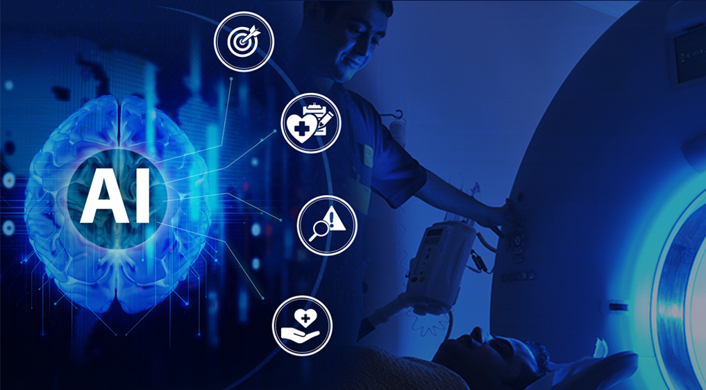
Taking a look at AI, we need to identify AI’s strengths in analyzing visual images. Researchers train the algorithms to better detect potentially dangerous abnormalities, generating faster and more accurate insights to help guide clinicians’ treatment decisions. AI adoption is sure to ease the overwhelming workloads impeding the profession, facilitating radiologists to do what they’re best at and perform them better.

Going further ahead, we can examine Workflow orchestration technology too. This promises to boost efficiency and alleviate bottlenecks. By directing cases to the right recipient in the correct order, this technology optimizes the effectiveness of the read, especially in teleradiology settings. With the profession’s ever-increasing need for solutions that match demand with supply, a lot of organizations provide solutions that facilitate better collaboration across facilities for effective workflow orchestration.

Teleradiology is another field that is assisting well where streamlining workloads is concerned. Remote radiologic coverage and reliable telecom infrastructures means more radiologic analysis is being performed online to take care of workloads between hospitals. And as the field becomes progressively digitized, apprehensions regarding the security of radiology data accentuate the need for robust solutions that will not just prevent breaches but at the same time also safeguard patient information while complying with regulatory requirements.

Diagnostic images captured at the right place and at the right time give physicians, surgeons, and care centers an important tool to help provide better patient care and at a reasonable cost. For this reason, Telerad Tech has been building out solutions since 2009.

Telerad Tech was established with the goal of optimizing radiology productivity and improving patient outcome delivery through transformational medical imaging software solutions. Today, it is amongst the market leaders in providing integrated RIS-PACS software solutions for teleradiology, medical imaging centres, and hospitals of all sizes globally. Telerad Tech’s solutions cater to workflows needs across departments, including Radiology, Cardiology, Podiatry, Orthopedic, Chiropractic, Oncology and Veterinary.

We are today amongst the market leaders in providing RIS with integrated PACS with significant installations in both cloud and enterprise environment across 1500 facilities in 24 countries.

Our software solutions suite has been incubated, tested and perfected in a radiology ecosystem and are designed to address the unique needs of multiple care pathways across departments, including radiology, cardiology, dentistry, oncology, and veterinary. Our software has customizable workflow features, intelligent productivity tools & analytics and Vendor-Neutral Archive technology. It has strong patient security framework and integrates seamlessly with other systems for exchange and retrieval of electronic health information.

To enable physicians to consistently deliver optimal patient management and to augment the precious time of radiologists, Telerad Tech has also leveraged Artificial Intelligence (AI), for various radiology diagnostics.

We truly believe that the future is here

Technological development has undoubtedly prompted some anxiety among radiologists. But while tech adoption will inevitably alter the way radiologists work, technology’s clinical value will be in supplementing and adding to and not replacing or even displacing the professionals. Radiologists empowered by AI will only encounter a new, more efficient stage of radiology, helping to focus their time and attention on the most crucial elements of their job.

Also, we need to remember that image analysis is just one of the aspects of a radiologist’s job, other tasks, including discrepancy reviews, diagnostic reasoning, and patient-facing work such as invasive radiology, will still be performed by humans. Those tasks will simply be supported and enhanced by advancing technology.

The future of radiology is here, and the prediction clearly states that it will not only better health care but also the lives of all the stakeholders.