Advances in the Teleradiology Sector

Advances in the Teleradiology Sector

Dr Arjun Kalyanpur, MD, Chief Radiologist and CEO, Teleradiology Solutions, Bengaluru, India speaks about the trends and prospects that teleradiology brings to healthcare

The last decade has been a truly exciting one for the teleradiology sector, which in its currently established form is about two decades old, i.e. as old as the current millennium. The first decade of this millennium had teleradiology proving its credibility as a potentially transformational technology innovation that could provide much needed support to radiologist-starved hospitals and diagnostic centres, emergency departments and perhaps most importantly, to other overworked radiologists. Over the second decade 2010-2019, however, teleradiology has seen exponential scale and has evolved into an industry unto itself. The main advances and trends facilitating this growth of teleradiology, which we have witnessed through the perspective of our own global clinical teleradiology practice have been:

  • Increase in utilisation of teleradiology – Once the initial concerns and doubts about its efficacy and safety were addressed, the sheer need for teleradiology based on critical radiologist shortages and dramatic increases in utilisation of imaging have driven its explosive growth. In our practise we have seen that the growth has spanned all geographies from the most technologically advanced metropolitan cities of the United States to remote and backward parts of Africa and Asia where healthcare delivery is still primitive. Similarly, all modalities have been impacted by teleradiology from plain X-rays to PET-CTs. And market surveys which a decade ago tentatively hinted at teleradiology growing into a 1-billion-dollar industry today seem to confidently proclaim that it will soon cross 10 times that number.
  • Clinical applications driving the need for teleradiology – Driving the demand for teleradiology has been a profusion of clinical applications in which teleradiology makes significant impact.
    • A classic example is acute stroke. In our US clinical teleradiology practice we have seen a significant increase in both the number of centres accredited as stroke treatment centres and the corresponding number of stroke imaging scans being performed (now including CT angiography and perfusion imaging). This means that thousands of images per patient need to be interpreted in an ultrashort time frame, typically within 10 minutes. Further, trauma and emergency imaging in general are increasingly utilised. In the US it is estimated that CT imaging of the spine for trauma increased by 400 per cent from 2006-2016 and Vascular CT angiography by 300 per cent over the same time period. As after-hours radiologists are hard to find in the current environment, teleradiology steps in as the provider of choice.
    • At the opposite end of the urgency spectrum even screening examinations such as chest X-ray for TB, low dose screening CT for lung cancer detection and mammography are able to realise greater adoption when combined with teleradiology interpretation. In parallel, the radiologist shortages have become even more acute, despite efforts to train more personnel, which are thwarted by the challenge that today radiologists in many instances are too busy with clinical work to be able to dedicate sufficient time to train the next generation. Hence, more teleradiology utilisation (and online education to train radiologists, but that’s another story…).
  • Cloud technology promotes the growth of teleradiology – From a technology standpoint one of the transformational trends in teleradiology over the past decade has been the spread of cloud based technologies which allows doctors and medical centres to set up and use teleradiology without investing in expensive hardware. Thus, there is a cost reduction by reducing the need for high-end onsite servers. It has also led to greater saleability of teleradiology as the ability to add on server space incrementally, rather than in quantum mode, is facilitated by cloud technologies. Also as server and internet bandwidth costs have dropped, the ability to reliably archive data offsite has become a reality. Rapid/immediate deployment of teleradiology services – as in the pay-as-you-grow model, such as the one afforded by our technology workflow Radspa, wherein any radiologist wishing to practice teleradiology can sign up for and switch on instantly, has accelerated adoption.
  • Teleradiology increases radiologist efficiency with workflow tools, viewer embellishments – Over the past decade the focus of teleradiology has been on further improving the reporting efficiency of radiologists by using an amalgamation of efficient viewer design, structured reporting, voice recognition and radiology lexicons all of which are driven by the increased workloads and radiologist shortages, a challenging scenario in which teleradiology provides its greatest value. Teleradiology workflows focus on ensuring that every second of radiologist time is optimally utilised.
  • Teleradiology enables working from home, part time: New service and business models have emerged in the past decade due to the proliferation of teleradiology services. The number of radiologists working part-time from home offices has increased in geometric progression which allows for better utilisation of that exceptionally valuable resource, that is radiologist time. This has also allowed for segments of the workforce who were previously unproductive such as young mothers and those dealing with elder care issues to remain productive in the workforce and equally importantly to retain their radiology skills. Teleradiology also facilitates a per-read reimbursement model that translates into more efficiency and greater productivity in reporting than the traditional salaried reimbursement model.
  • Recent governmental adoption and tendering process for teleradiology – A significant advance in teleradiology over the past decade has been a consequence of the realisation on the part of the government that it is unable to effectively address on its own the radiology needs of the community and its embracing public private partnership models which use teleradiology workflows to deliver valuable access to remote areas in a public health environment. Our work with the government of Tripura has illustrated the power of collaboration between the private sector and government using teleradiology as a bridge to provide radiologist access to district hospitals and community health centres to rural populations in remote parts of India.
  • 3D Teleradiology Lab – the use of outsourced 3D post-processing services for generation of high quality volume data that facilitates surgical planning is an important offshoot of teleradiology that has been firmly established over the past decade. Our work with the Harvard MGH 3D Lab providing after hours post-processing services to a luminary tertiary care metropolitan hospital site is an illustration of the value of this service. From the technology standpoint, the convergence of 3D workstations with teleradiology workflow is a new advance that increases and enhances the reach and value of both.
  • Teleradiology core lab facilitates drug discovery and clinical research – In the last decade, there has been a resurgence in drug discovery related research in the pharma and biotech sector in the country while the industry has only continued to grow at a global level. The use of teleradiology-driven Core Labs, such as our own Image Core Lab, by the pharma/biotech sector has significantly increased, as has the creation/adoption of teleradiology workflow such as Clinspa which are focussed on the unique requirements of clinical research-related radiology reporting such as dual read with adjudication, quantitative imaging and the use of objective reporting standards such as RECIST. Teleradiology has proven itself to be a major value addition in drug discovery and clinical research.
  • Teleradiology-driven education and second opinion sites– The increasing use of teleradiology for second opinions and subspecialty consultations especially through web-based services to which anyone can upload their imaging data marks another success story for teleradiology in recent times. And the growth in popularity of teleradiology-driven online education websites portals for radiology education parallels that of the Internet as the foremost source of information and learning today. We currently have users from all across the world logging on to our live e-lectures by luminary international guest speakers.
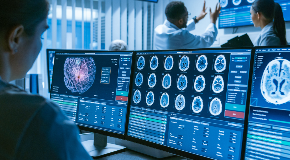
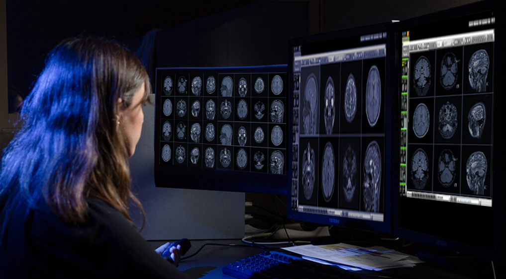
  • Teleradiology and AI, the next big thing – Of all these, by far the most exciting development in the teleradiology space has been the emergence and realisation of growing synergies between it and the new and precocious kid on the block, namely artificial intelligence and its conjoined twin, deep learning. Both teleradiology and AI thrive in the environment of clinical complexity, computational and networking technology and big data and both seek to solve the same fundamental challenge of too many images and not enough radiologists. Teleradiology provides an exceptional environment for the development, validation, testing and finally the deployment of deep learning algorithms. The integration of AI algorithms with teleradiology workflows can help make the radiologist more efficient and more accurate. For instance, we have deployed an AI tool that can auto detect a brain bleed into our ER teleradiology practice which enables stroke cases to be triaged immediately as positive or negative. This represents a pivotal moment in the evolution of the new age technology enhanced super-radiologist. AI is the catapult that will propel teleradiology into the new decade and beyond.

To summarise, from a teleradiology perspective, very exciting times lie ahead… and to paraphrase the old time jazz singer Al Jolson…. we ain’t seen nothing yet!

References:

1. https://www.mpo-mag.com/contents/view_breaking-news/2019-08-29/teleradiology-market-continues-to-grow/
2. https://www.auntminnie.com/ index.aspx?sec=rca&sub=rsna_2018&pag=dis&ItemID=123892
3. https://www.diagnosticimaging.com/ articles/cloud-computing-creates-climate-change-teleradiology
4. https://www.dicomstandard.org/wp-content/uploads/2018/10/Day3_S2-Bedi-Clinical-Teleradiology-Workflow.pdf
5. https://www.rcr.ac.uk/posts/nhs-does-not-have-enough-radiologists-keep-patients-safe-say-three-four-hospital-imaging
6. https://www.slideshare.net/ TeleradiologySolutio/teleradiology-in-the-northeast
7. https://health.economictimes.indiatimes.com/news/medical-devices/teleradiology-solutions-invests-300000-for-3d-imaging-facility-partners-with-massachusetts-general-hospital/54419957
8. https://radiologykey.com/evaluating-and-working-with-an-imaging-core-laboratory/
9. http://www.arjunkalyanpur.com/2018/10/elearning-from-very-best-in-world.html
10. https://www.researchgate.net/publication/333411163_Teleradiology_and_Artificial_Intelligence_-_Birds_of_the_Same_Feather

Artificial Intelligence all set to Virtualize Radiology Brains

Artificial Intelligence all set to Virtualize Radiology Brains

Mr. Prashant Akhawat’s interview with Live Mint

  • AI and automation will not replace radiologists, but will act as an assistant to radiologist in every step of the imaging detection, diagnosis and prognosis
  • There is an increase in interest and enthusiasm for AI among the radiology community as the discussion is moving upwards on from considering AI as a threat
Accelerating with an exponential growth, artificial intelligence (AI) is all set to move from experimental stages to live industry implementations and all is set to mark its presence across all industry verticals. AI is all about virtualizing human cognitive functions in the form of software brains. For organizations, harnessing AI is not optional, albeit it is critical to stay competitive. Gartner in its recent study (2018), predicts the business value derived from AI to reach $3.9 trillion by 2022. With the disruptive potential, the investments in AI are ever-increasing. It is redefining industries with automation processes, and personalization. The healthcare industry has been one of the foremost adopters of the AI amongst all others. The advancement, the healthcare industry is reaping with the use of AI to maintain medical records, do mundane tasks more accurately and faster, to design care pathways, digital consultations, medication management, drug creation, image analytics, bigdata analytics, medical robotics, health monitoring and host of other things.

Powering Radiology with AI

Today, in India, there is a growing demand for improved medical care. We see an increase in insurance penetration, the rise in chronic disease and an aging population — all this demand for better imaging diagnoses and treatment. Radiology industry is facing its own set of ongoing challenges – the shortage of radiologists topping the list. The radiology industry is a functional domain for automation by virtualizing domain intelligence into an intelligent software. The quantum leap in medical imaging technology has led to exponential growth of medical imaging data stored digitally. Deep Learning algorithms and Image Analytics can help in improving medical diagnosis and aid radiologists with better reporting efficiencies.

Nowadays, with the growing incidence of lifestyle diseases, the requirement of frequent imaging and getting multiple scans at a higher resolution has increased. There are scenarios where the hospitals have X-Rays, MG, CT and MRI equipment’s but sans radiologist to read a report. To bridge the gap where AI can play a pivotal role by performing specific tasks such as image recognition – nodule detection, hemorrhagic or ischemic stroke detection, fracture detection, breast cancer analysis with prior case analysis and other narrow tasks requisite identify potential findings in medical images, which is one set of tasks performed by radiologists. This gives a window to the radiologists to focus more on image-guided medical interventions, defining clinical parameters of imaging examinations, relating findings from images with medical records and test reports, consult physicians for treatment based on the diagnosis, discussing procedures and results with patients. Artificial Intelligence is taking over image reading and interpretation so that radiologists can read more images in a short span with better accuracy as the number of images has increased more in the last decade than the number of radiologists.

Radiologists need to adapt themselves to new skill sets and technologies for attaining better productivity by integrating AI with radiology practice. AI and automation will not replace radiologists, but will act as an assistant to radiologist in every step of the imaging detection, diagnosis and prognosis and will also help in prior analysis and comparison. AI Algorithms will have a stronghold in medical imaging and will become an integral part of RIS-PACS, as frequently we hear some or the other algorithm is developed to detect tumors, lesions, fractures and host of other things.

With the help of RIS-PACS workflow enabled with AI, a radiologist sitting at any location can do the reads for any healthcare center located remotely, which addresses the accessibility challenges, subspecialty reads, point in time care with reduced costs. Further helping them to optimize results without compromising on the accuracy of diagnosis. For emergency reporting (trauma and stroke cases) AI can assist the radiologist with a preliminary report with which they can conclude with their final interpretation of the case within the stipulated time. AI Enabled RIS-PACS can prioritize stroke or stat cases and reduces turnaround time in turn will improve patient care. The algorithms are trained to assist the radiologists with various detections, diagnosis, staging, sub classification of different medical conditions. Hence, deep learning is a shoulder to the increasing workload in radiology.

Global Market for AI

Artificial intelligence’s complexity is not a deterrent to its adoption instead it is as disruptive as the internet was. The future of investment is AI, and it has not just been observed, but also proved that AI helps in improving the radiologist’s productivity, efficiency and quality diagnosis, hence this would help them reduce costs and increase return on investments. In the healthcare industry, AI is rapidly rising in the medical imaging domain. Globally, the AI market in medical imaging is forecasted topping US$ 2 billion by 2023. Notably, there is an increase in interest and enthusiasm for AI among the radiology community as the discussion is moving upwards on from considering AI as a threat. Also, clinical applications have shown improved clinical results with the use of AI.

All the research trends underline how AI is revolutionizing radiology in the long run. AI-based companies have learned to warm-up radiologists. Realizing the technological potential of AI, radiology practitioners are partnering with AI ventures to have a seamless RIS-PACS workflow. AI is going to augment the way care is provided by healthcare practitioners.

Picture Archiving and Communication System (PACS) and Its Benefits

Picture Archiving and Communication System (PACS) and Its Benefits

Technology and innovation play a major role in today’s healthcare system as it is crucial for sustaining health. It has enhanced the quality of medical care offered to patients, and PACS system in hospitals & health facilities is one such example of technology improving medical care.

PACS (Picture Archiving and Communication System) has changed the way radiology works and is now considered one of the most essential requirements in healthcare facilities. Sharing of instant medical images electronically and reporting them remotely is now very easy and quick, thanks to this invaluable software. With AI (Artificial Intelligence) enabled technologies now becoming available in PACS, their functionality is growing by the day

What is PACS?

PACS (Picture Archiving and Communication System) is a medical imaging technology that provides economical storage, presentation, retrieval, distribution, and management of medical images. Transmission of electronic images and reports takes place digitally via PACS. Thus, manual filing, retrieving and distribution of film jackets is no longer required. It allows storage and viewing of all types of medical imaging by healthcare organizations both internally and externally.

A radiology PACS is often deployed with RIS. An RIS is used to record patient history and schedule appointments, whereas PACS focuses on image storage and retrieval.

The four major components of PACS are:

  • The imaging modalities
  • Transmission of patient information through a secured network
  • Interpreting and reviewing of images through a workstation
  • Storage archives for retrieval of images and patient reports

Benefits of PACS

PACS improves efficiency in electronic data handling workflow. It offers a cost and space advantage due to decreasing price of digital storage. Benefits provided by PACS are many, but here we highlight few of the most important ones:

  • Improved Viewing and Analysis

    An effective viewing and analysis is possible as PACS’ digital images enable you to zoom in and operate the images for a more elaborate analysis.

    Where the conventional film can only exist in one place at one time, PACS enables simultaneous multi location viewing of images. It enables collaboration among radiologists as they can seek each other’s opinions by viewing the cases simultaneously and discussing them under peer review module. The interpretive skills of the professionals prove beneficial for the patients as well. The high-quality images make it possible to give a more accurate diagnosis.

  • Easy Accessibility to Images and Reports

    PACS enables instant and easy access to images and reports. No matter where the tests are performed reporting can be done remotely and results can be shared anywhere, even if it is an isolated facility. PACS enables submitting reports, archiving images and transferring them through a portable media anywhere in the world. Practitioners at different physical locations can access the same information concurrently for teleradiology. In addition, quick access to prior images is also possible at the same institution. Radiology history of patients is available, which allows comparison with previous studies.

  • User-friendly Software

    User-friendly since there are several customizations available for easy use of the software for staff and beneficial for patients. PACS is a great integration platform for other automation systems such as Radiology Information System (RIS), Electronic Medical Record (EMR), and Hospital Information System (HIS). The PACS database automatically groups all images chronologically, correctly labelled according to their examination. They can also be retrieved easily with the help of criteria’s such as name, hospital, referring clinician, etc.

  • Efficient Data Management

    The system provides an efficient, seamless review of radiology cases within a physician’s daily workflow as it makes it easier to store and organize imaging data, with a centralized and accessible system. Data management becomes more efficient as the number of duplicate images can be reduced as previous data is available with the system.

Steps to Consider While Purchasing or Upgrading PACS

While purchasing or upgrading to a new PACS, one should look for features such as scalability and user-friendliness. The features should be easily configurable and should have uploading features for prior studies. Other than that, it should have a voice recording feature, integration with the Hospital Information System (HIS) and the Cloud feature. It is also beneficial switching to a new system when it has in-built AI algorithms.

An integrated RIS-PACS is a great advantage to radiology department as it can give evidence-based insights such as enhanced productivity of radiologist, turn-around time, modalities being used for improved workflow, and referring physicians sending maximum exams. RADSpa, one of the best integrated RIS-PACS available, provides a customizable work-flow suiting the requirements of radiologists.

A RIS-PACS’ rich intuitive AI integrated workflow, supporting all DICOM modalities, an orchestrated work-flow, multilingual software, advanced application supporting MIP/MPR and 2D/3D viewer, a scalable architecture and ability to integrate with any existing PACS and 3rd party AI algorithm, is sure to add value and increase productivity for any radiology department.

Artificial Intelligence in Emergency Radiology

Artificial Intelligence in Emergency Radiology

A Report, based on the Keynote Speech delivered
By Dr. Anjali Agrawal, Head, Teleradiology Solutions, Delhi Operations
@ Artificial Intelligence in Radiology 2018 Symposium, November 10, 2018
Organized by Telerad Tech and Image Core Lab

Dr. Anjali Agrawal addressed the audience about how AI was going to be relevant in emergency care and emergency radiology.

Deep learning and the human brain

Speaking broadly on artificial intelligence (AI), Dr Anjali said that AI is a more gen­­­eral term and includes machine learning (ML) and deep learning (DL). Machine learning, a specific type of AI, gives computers the ability to learn without being explicitly programmed. Deep learning, a subset of machine learning, mimics the human brain configuration, where the multiple neuronal layers or neurons can crunch vast amounts of data and draw conclusions. In particular, DL has immense relevance for radiology and healthcare. The availability of large amounts of annotated image datasets and increased computational power had made AI a reality and it wasn’t an illusion anymore. It is moving from experimental to the implementation phase now.

Current state of Emergency and Trauma Care

Dr. Anjali drew attention to the current state of emergency and trauma care in India. From trauma registry, it was a documented fact that trauma related deaths in India occurred every 1.9 minutes. The mortality in serious injuries was 6 times worse in a developing country such as India, as compared to a developed country. A WHO survey revealed that there were more deaths due to lack of timely care than due to other diseases like AIDS, Malaria and TB, all put together.

Dr. Anjali said that, more than 80% of Indians didn’t get care within the golden hour and she highlighted the challenges in emergency care and radiology. There was tremendous pressure on the limited resources that were available at one’s disposal. The process from the scene of accident to the emergency room is disorganized. Dr. Anjali drew attention to the education system and training, which was quite heterogeneous.

AI will transform the ER services

As per Dr. Anjali, AI could be very useful for triage in the emergency room. Studies have shown that Emergency Severity Index assignment by doctors and nurses is correct only 60% of the time. They ended up under-triaging almost 27% of the patients. And a vast majority of those, nearly half, went into the mid-acuity group, level 3 – a typical human tendency to play safe.

AI and deep learning could help by analyzing the complex data from various sources – the age and sex of the patient, presenting history, complaints, vital signs, what was the mode of transport – did the patient walk-in or was s/he brought by an ambulance, past medical history etc. This could help in transforming the emergency department operations. By using these algorithms one could accurately triage patients, so that the critically ill patients got the attention of the emergency medicine physician and were managed appropriately. These algorithms could also help in allocating resources appropriately, minimizing mismatch between staff and patient case load, with improved patient outcomes. AI could help expedite interpretation of emergent imaging studies. These algorithms would be able to make predictions of adverse events and help make an individual-specific follow-up plan.

AI will revolutionize the radiology workflow and create smart enterprises

Dr. Anjali maintained that disruptive new generation healthcare technologies such as AI, robotics, machine learning and deep learning will revolutionize radiology workflow in many ways and that it was going to lead to massive improvements in quality, value and the depth of contribution of radiology towards patient care. She stated that one of the most well researched applications is emergency radiology.

Dr. Anjali cited that, AI could help make an informed decision regarding the need for imaging and the choice of modality based on analysis of the patient records. AI enabled algorithms could play a huge role in reducing radiation doses of CT examinations or reducing scan time for MRI, by using various enhancement and post-processing techniques. However, the overall decision making and communication with the referring physician or the patient would require the intervention of a human radiologist for quite some time, despite being aided by AI.

AI can help reduce scan timings and dosages

Dr. Anjali said that, the algorithms could enhance very noisy, grainy and undersampled data, such as from MRI, which were being produced in shortened timeframes and produce high-resolution MRI images– with huge implications in the emergency room, where one tends to shy away from doing an MRI because of time constraints. For e.g., if an MRI could be done in two- thirds or one-third the time required, one would be more comfortable in sending a sick patient for a suspected hip fracture for an MRI.

Similarly, these algorithms could also be applied to Computed Tomography (CT) scanners to help reduce the CT radiation dose – a huge advantage, as the reduction in radiation dose from CT would be comparable to a standard chest x-ray. One would be able to do ultra low-dose CTs, and get more information from diagnostic quality images, compared to a radiograph.

AI can ease the workflow of a radiologist in many ways

Showcasing a typical workflow of a radiologist, Dr. Anjali said that the radiologist logs into the system, reviews his/her work list and selects a study to review. The radiologist reviews other information such as patient history, prescriptions, etc., that are related to the case. Once the images are presented, the radiologist, in most cases, adjusts the hanging protocols to enable him/her to perform the interpretation and generate a report. The initial process of arranging studies is time consuming. Citing the recent developments in reading protocols, Dr. Anjali said that it could help hang the images in an interactive manner, learning each time, catering to individual preferences, and saving time.

AI can help in detection of findings, segmentation, quantification and reporting, in a manner that is easier to understand by both the referring physicians and the patients.

Dr. Anjali maintained that AI could help the radiologist by triaging cases, such that only the positive ones could be seen by the radiologists for further interpretation. She was of the thought that, as opposed to a few articles quoting that AI would replace a radiologist for particular targeted applications,  AI would assist a radiologist, where a radiologist would act as a second reader or vice versa.

She gave one particular example where an article looked into automated detection of critical findings on non-contrast CT examinations of the head –hemorrhage, mass effect, and hydrocephalus using an AI algorithm, which would be helpful in triage. If the algorithm found the non-contrast CT to be negative, it went through another stroke algorithm. In the case of it being positive, it was labeled as a critical imaging finding, and if they were both negative, it was labelled as “no critical imaging finding”. The algorithm had a good sensitivity of 62% and a specificity of 96%, comparable to a radiologist in the detection of acute ischemia. The sensitivity and specificity were higher for detection of hemorrhage, hydrocephalus and mass effect, matching the performance of the radiologists. Therefore, the study concluded that there was a huge potential for AI algorithm in terms of screening and detection of critical findings in the emergency setting.

Dr. Anjali said that she had seen similar data in her group and had detected intracranial hemorrhage with a very high sensitivity and specificity using a hybrid approach of convoluted neural networks and factorial image analysis.  The data and the results were comparable to the existing literature and that more data pertaining to quantification and localization of intracranial hemorrhage is underway.

Juxtaposing her earlier statement on triage being the low hanging fruit for AI applications in radiology, Dr. Anjali, mentioned that apart from acute neurologic conditions, these triage tools had been used in the detection of chest radiographic findings by classifying them into normal or abnormal with a high accuracy of almost 95%.

She quoted another example of AI application – wrist fractures. These algorithms were trained by senior orthopedic surgeons. When the emergency room physicians, not trained orthopedicians or radiologists, used them, their sensitivity improved from 81% to almost 92% and specificity from 88% to 94%; with a relative reduction in misinterpretation rate of almost 47%. Dr. Anjali simplified it further by saying that the algorithm was able to emulate the diagnostic acumen of the experts by providing the labels on where the fracture was and also put a heat map, assigning a confidence level to the detected fracture.

Adding on, Dr. Anjali said that AI applications would also be useful in detection of non-acute findings the emergency radiology setting. These may be overlooked because the focus is on the critical life-threatening illnesses. AI could help with measurements of bone density, detection of fatty liver, coronary calcifications, and the presence of emphysema, which may not be relevant in the acute setting, but would have implications in the future.

AI can make expertise widely available and scalable

Dr. Anjali then stated that according to her, AI would become a huge leveler in terms of the expertise and experience of radiologists. Good radiology consult would become easily accessible, affordable, as well as scalable. In the scenario of a mass casualty incident, the algorithms could be put to use to quickly distinguish between critical and non-critical cases. Handling massive imaging volumes would also become easier.

Dr. Anjali mentioned about one particular study on automated bone age estimation where the algorithms were extremely accurate as well as reproducible, with an interpretation time of fewer than 2 seconds. This is a huge achievement because every radiologist knows how tedious and time-consuming the task of bone age determination is.

Man with machine synergy

She went to add that many similarities had been drawn between the fields of medicine and aviation. The pilot, as well as the doctor, needs to be highly skilled, as they are responsible for human lives. Both the professions have benefitted tremendously from automation. There is no flight without a human pilot, and similarly, there would be no healthcare without human doctors, because the legal responsibility would always be with the doctors. Dr. Anjali urged the doctors to not forget that medicine was an art and that the physicians needed to practice it like an art to stay relevant. AI would not replace radiologists. According to Dr. Anjali, it would be the synergy between man and machine that will help the profession as well as benefit the patients.

Artificial Intelligence in Teleradiology

Artificial Intelligence in Teleradiology

A Report, based on the Keynote Speech delivered
By Dr. Arjun Kalyanpur, Chief Radiologist and CEO, Teleradiology Solutions
at Artificial Intelligence in Radiology 2018 Symposium, November 10, 2018
Organized by Telerad Tech and Image Core Lab

Dr Arjun Kalyanpur began his speech recalling how Teleradiology Solutions for the past few years has been engaged in the field of artificial intelligence (AI) with the health-IT company Telerad Tech and drug trial company Image Core Lab. He also congratulated Telerad Tech for launching MammoAssist – its AI software for breast cancer detection and also for establishing a new dedicated AI lab facility at the same time.

Dr. Kalyanpur noted that his company Teleradiology Solutions had reached an inflection point in its journey in the direction of artificial intelligence and it was an opportune moment for their company to be part of an event like this and be amongst the distinguished speakers in the field of AI.

AI and Teleradiology – synergistic and disruptive technologies

As per Dr. Kalyanpur, both AI and Teleradiology are very closely linked to each other and are extremely synergistic. They both address the same fundamental clinical issues about shortages of radiologists and both provide technology-enabled solutions for the same clinical issues.

Revisiting their journey of how teleradiology brought them to the current point where AI is seen as the direction forward, Dr. Kalyanpur said that the drivers that set them on this path 15 /16 years ago was the shortages of radiologists with most of them being focused in metros, and emergency radiology being the most severely affected area and still continues being one today.

He noted, that the numbers of radiologists haven’t changed much in the past 20 years and that there was still a dramatic shortage of radiologists the world over. Drawing up a scenario, Dr. Kalyanpur highlighted that even a developed country like the US with the best radiologist to population ratio, considered itself of being in the deficit with India being in the middle of the curve somewhere, whereas Tanzania on the other end had 1 radiologist for over 1 million population.

AI is important for addressing the increasing burden of diseases

He cautioned that, there is an increase in chronic diseases, due to progressively unhealthy lifestyles and living conditions, all of which required repeated imaging at multiple times in a patient’s life, leading to an increase in the number of images and their complexity in studying them. He added that in the 123 years, between the first X-Ray being produced in Roentgen’s Lab, till today, advancements have led to a dizzying array of imaging modalities of extremely high resolution, in all shades and hues being available to the radiologists, increasing work for them analyzing the data.

Understanding the gaps, Dr. Kalyanpur noted that, 15 years ago teleradiology evolved as a solution to these factors delivering the value proposition of not needing to have a radiologist at every hospital. It was technology driven solution with work flow and efficiency being at the core. It also had a quality benefit because the images were being brought to the most qualified radiologist, spawning the term “nighthawk”. The company was serendipitously at the forefront in the sector, providing night shift emergency services to hospitals at different parts of the world resulting in better performance and better quality services.

Emphasizing again, Dr. Kalyanpur said that, both teleradiology and AI were essentially disruptive technologies. Having been through and seen the evolution of teleradiology, he felt that, AI was seeing the same disruption as they had witnessed in teleradiology. Citing similarities between the two, he noted, that the resistance pattern was similar, and AI also faced the same concerns and fears that teleradiology had previously faced. Depending on how one would see them, both could either be seen as a threat or as a benefit. The same drivers were present for both innovations. Since both are IT enabled solutions and follow Moore’s law principles, one could only expect them to be faster, more effective and cheaper in the coming years. He also noted that both had the potential to transform healthcare by simultaneously increasing radiologist quality and productivity – a double whammy in terms of their benefit and impact on healthcare.

Key trends in radiology

Describing radiology as a quantitative science, Dr. Kalyanpur said that radiology today was beyond a simple image description. Studying a few trends in radiology prevailing today, Dr. Kalyanpur chose to highlight 3 of them, sub-specialization being one. Further, he noted that there was an increase in focus on report turn-around time as all of medicine was becoming metrics- driven. Turnaround time was becoming the basis of judging the performance of a radiologist or a radiology group. He also pointed out that the evolution of cloud-based workflows also had made a tremendous impact in the field.

Citing stroke as an example, Dr. Arjun said that, in the 15 years that they had been practicing teleradiology the specialty had evolved to the point where it was no longer just a matter of looking at the CAT scan of the brain and determining whether it was a stroke or not. Today they had a CT angiogram or a perfusion CT to analyze as well, increasing the number of imaging modalities available to assist in clinical decision making. Quantitative imaging today helped in quantifying the size of the stroke, determine the collateralization of the brain and the impact on patient outcome. Therefore, radiology was no longer just a diagnostic science but had evolved into being a prognostic tool while Quantitative imaging was available in every sphere – Whether it was tumor detection, tumor analysis or quantification of vascular disease.

Looking at it from the operational standpoint, Dr. Kalyanpur noted that turnaround time was another factor that entities needed to be conscious of. He mentioned that, every tele-radiologist in the organisation practiced medicine under the scrutiny of the stopwatch. Citing it as a challenge, he said that, finding a fine balance between quality and speed was important as the faster a radiologist reads an image, the greater was the chance of him/her missing a finding. As per him, teleradiology had made the important aspect of work flow possible by allowing decentralized reporting and being a cloud-based solution, a radiologist could read for hospitals in any part of the world, with the understanding that compliance with licensure and certification was adhered too.

Making a radiologist more efficient and productive

Taking a step back and speaking on his role as a radiologist, Dr. Kalyanpur said that, usually a radiologist detected and quantified the lesion, analyzed the finding, communicated and recommended the follow up. Tackling the criteria on making the radiologist more efficient and productive, he suggested making the process of reporting, the work flow smooth by providing all the relevant information to the radiologist and assisting them by creating a templated report. Speaking on the productivity, Dr. Kalyanpur said that, the average productivity of a radiologist in the world was between 6-10 RVUs (relative value unit) an hour. That could probably be increased by about 15-20% by using these techniques. However, AI could potentially double the productivity of a radiologist, thereby helping in solve the radiologist shortage issue of half the world.

Highlighting the larger issue of radiologist errors, Dr. Kalyanpur mentioned that as per some studies, 3-5% of radiologist reports contain errors, such as for example small bleeds in the brain, small clots in the lungs or very subtle fractures in the spine. Considering that a billion scans were performed around the world every year, it meant that around 30 million scans would have reporting errors – a gravely concerning huge number. He opined that radiologist accuracy and patient outcome could be improved by allowing AI to detect these findings.

Predicting on the estimates on the size of the industry, Dr. Kalyanpur said that, the initial figures in teleradiology suggested it being a billion dollar industry, however it had grown beyond 4 billion currently and he was confident that it would further grow. Similarly, he was optimistic about AI growing beyond the current 2 billion in the future and wished best of luck to Telerad Tech for its endeavours.

Developing AI in radiology

Speaking on the importance of developing AI in radiology, Dr. Kalyanpur said that, it was important to have communication between radiologists, scientists and engineers. This would enable data with labelled information help in developing the tools and would require radiologists to validate them once they were developed to confirm that they actually worked. He suggested that this could be integrated into the workflow most effectively by integrating it into the teleradiology workflow which allowed these technologies to be deployed at scale across the globe, where the entire world population of radiologists could be the consumer.

The synergies between teleradiology and AI was that at every stage of the AI cycle teleradiology had a valuable role to play and offer – whether it was data acquisition from around the world (as many as over 200 hospitals in 20 countries) forming a huge repository for analysis, testing an algorithm in a teleradiology environment and the teleradiology workflow to be used as a distribution tool for AI software.

Simplifying it, Dr. Kalyanpur noted that, teleradiology could be seen as being a shopping mall, in which each AI algorithm was an individual store or teleradiology could be an I-phone on which each individual app resided and allowed the consumer to connect with the product. He expressed his satisfaction to note that Telerad Tech, apart from developing its own software algorithm, was also partnering with other entities in the same space. And allowed their workflow to be used as a distribution module for teleradiology algorithms or for AI algorithms to radiologists around the world allowing all possibilities of detection, quantification triage for smaller lesions and detection for the larger lesions.

In his concluding remarks, Dr. Kalyanpur said that, it was all about perspective and focus. Quoting radicals such as Professor Hinton, who opined that since AI would have taken over in the future, training of radiologists should be stopped because we wouldn’t need them anymore on one hand to moderates such as Professor Langlot, Stanford, on the other who believed that radiologists who used AI will replace radiologists who wouldn’t in the future. He suggested that it would be wiser to have a pragmatic approach since it was an evolutionary area. He was of the view that one needs to learn to adapt and understand the space so that one could utilize it better. Citing Darwin he wrapped up his speech by saying that only the most responsive to change would survive over the more stronger or the more intelligent.